
Glycobiology
RayMol integrates dedicated glycobiology tools directly into its molecular model and Metal rendering pipeline. Recognized carbohydrate residues can be displayed as three-dimensional Symbol Nomenclature for Glycans (SNFG) shapes or colored using the standard SNFG palette, while the underlying atomic coordinates remain fully interactive and inspectable.
SNFG conventions & residue catalogue
Section titled “SNFG conventions & residue catalogue”RayMol adheres to the Symbol Nomenclature for Glycans (SNFG) standard established by the glycomics community and NCBI. Carbohydrate residues are represented by standardized geometric shapes and colors based on their monosaccharide family.
The built-in catalogue covers the most common mammalian monosaccharides and their chemical-component PDB identifiers:
| PDB Code | Monosaccharide | SNFG Symbol | 3D Shape | Color | Hex Code |
|---|---|---|---|---|---|
NAG, NDG |
N-acetylglucosamine | GlcNAc | Cube | Blue | #0072BC |
A2G, NGA |
N-acetylgalactosamine | GalNAc | Cube | Yellow | #FFD400 |
BM3 |
N-acetylmannosamine | ManNAc | Cube | Green | #00A651 |
MAN, BMA |
Mannose | Man | Sphere | Green | #00A651 |
GAL, GLA |
Galactose | Gal | Sphere | Yellow | #FFD400 |
GLC, BGC |
Glucose | Glc | Sphere | Blue | #0072BC |
FUC, FUL |
Fucose | Fuc | Cone | Red | #ED1C24 |
SIA, SLB |
N-acetylneuraminic acid | Neu5Ac | Diamond | Purple | #A54399 |
NGC |
N-glycolylneuraminic acid | Neu5Gc | Diamond | Light Blue | #8FCCE9 |
Residue codes not in this table are left untouched; a catalogued residue whose ring atoms are incomplete gets no 3D symbol.
Rendering SNFG cartoons
Section titled “Rendering SNFG cartoons”When an SNFG cartoon is generated, RayMol computes the centroid and ring normal of each complete sugar ring, renders the corresponding 3D geometric symbol, and traces covalent glycosidic bonds between residues as cylindrical linkers. Linkers reflect true covalent connectivity, not arbitrary spatial proximity.
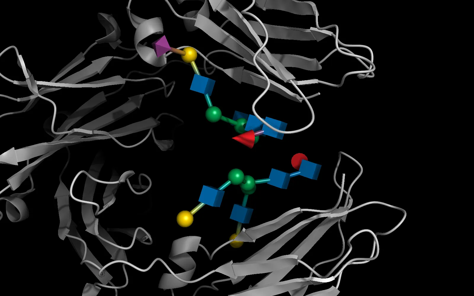 SNFG glycan cartoon of a sialylated IgG Fc (PDB 4BYH).
From the interface
Section titled “From the interface”In the Objects panel:
- Open the S menu on an object row and choose Glycan cartoon to generate the 3D symbols.
- Open the H menu on an object row and choose Glycan cartoon to remove its 3D symbols and show those sugar residues as sticks.
- Open the C menu on an object row and choose SNFG glycan palette to color the underlying atomic model according to SNFG standards without generating 3D cartoon shapes.
From the command line
Section titled “From the command line”Run glycocartoon and glycocolor directly in the command entry bar:
fetch 4byh, async=0glycocolor 4byhglycocartoon 4byhTo customize cartoon dimensions or linkers, supply the optional arguments:
glycocartoon [selection [, size [, draw_linkers [, state]]]]selection(default"all"): The target atom selection.size(default2.5): The geometric scale of the generated 3D symbols in angstroms.draw_linkers(default1): Set to1to render connecting cylinders along glycosidic bonds, or0to render only the monosaccharide symbols.state(default1): Coordinate state index for multi-state objects.
Hiding cartoons
Section titled “Hiding cartoons”To remove one cartoon, pass the same selection text you gave glycocartoon (e.g. glycocartoon_hide 4byh after glycocartoon 4byh). If unsure, use glycocartoon_hide_all.
glycocartoon_hide 4byhTo remove all generated glycan cartoon objects across the entire session and show those sugar residues as sticks:
glycocartoon_hide_allInspecting glycan topology & geometry
Section titled “Inspecting glycan topology & geometry”Complex N- and O-linked glycans form branched tree structures rooted at protein attachment sites (such as asparagine for N-glycosylation or serine/threonine for O-glycosylation).
The Glycan Topology panel
Section titled “The Glycan Topology panel”To inspect the glycan forest interactively:
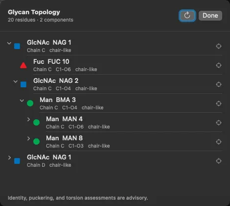 The Glycan Topology sheet for the IgG Fc N-glycans of 4BYH.
- Locate the OBJECTS section header in the Objects panel.
- Click the indented-list icon (Glycan topology) at the right of the OBJECTS header.
- The Glycan Topology sheet opens, displaying each glycan tree, rooted at the sugar covalently bonded to a non-sugar residue (for example the Asn of an N-glycan) when there is one. The header shows the residue and component counts.
Each node in the topology tree details:
- Residue: SNFG symbol, residue name, number and chain.
- Ring shape: a coarse puckering class (chair-like, boat/skew-like, envelope/half-chair-like, nearly planar).
- Linkage: the bond atoms (e.g. C1-O4).
Numeric Cremer–Pople Q/θ/φ and glycosidic φ/ψ (and ω for 1→6 linkages) are available from the glyco_tree command. Clicking any node in the topology list highlights and centers that residue in the 3D viewport.
Topology commands
Section titled “Topology commands”The same structural analysis can be invoked from the command bar or Python scripts:
glyco_tree [selection [, state]]glyco_remediate [selection [, state]]glyco_tree: Constructs the covalent glycan tree hierarchy, measures ring puckering parameters, calculates glycosidic torsion angles, and prints the result as JSON (residues, Cremer–Pople Q/θ/φ, linkage φ/ψ/ω).glyco_remediate: Reports, for each residue in the selection, whether its residue code is in RayMol’s SNFG catalogue. For unknown codes it says whether the atom names suggest an aldose-like or sialic-acid-like ring. Nothing is changed; output is JSON.